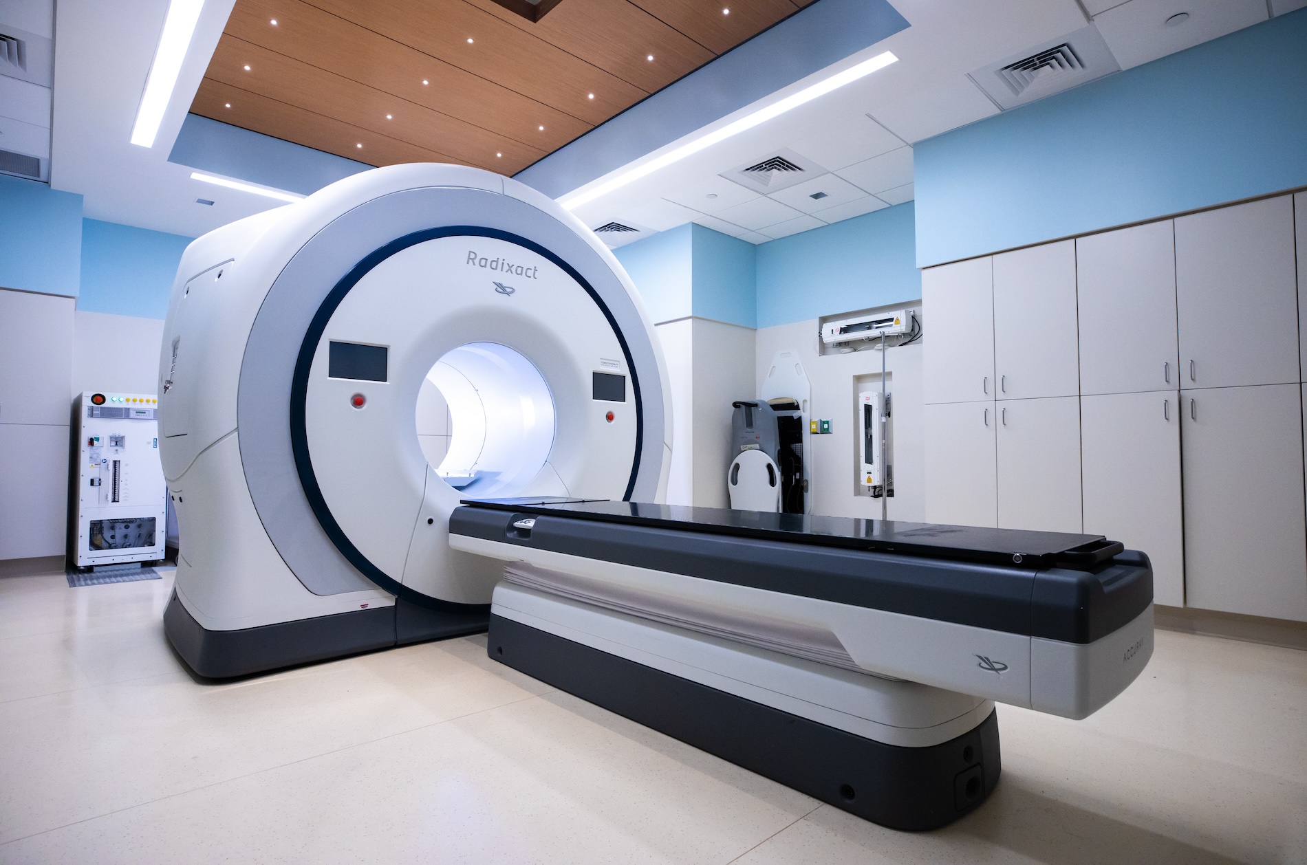
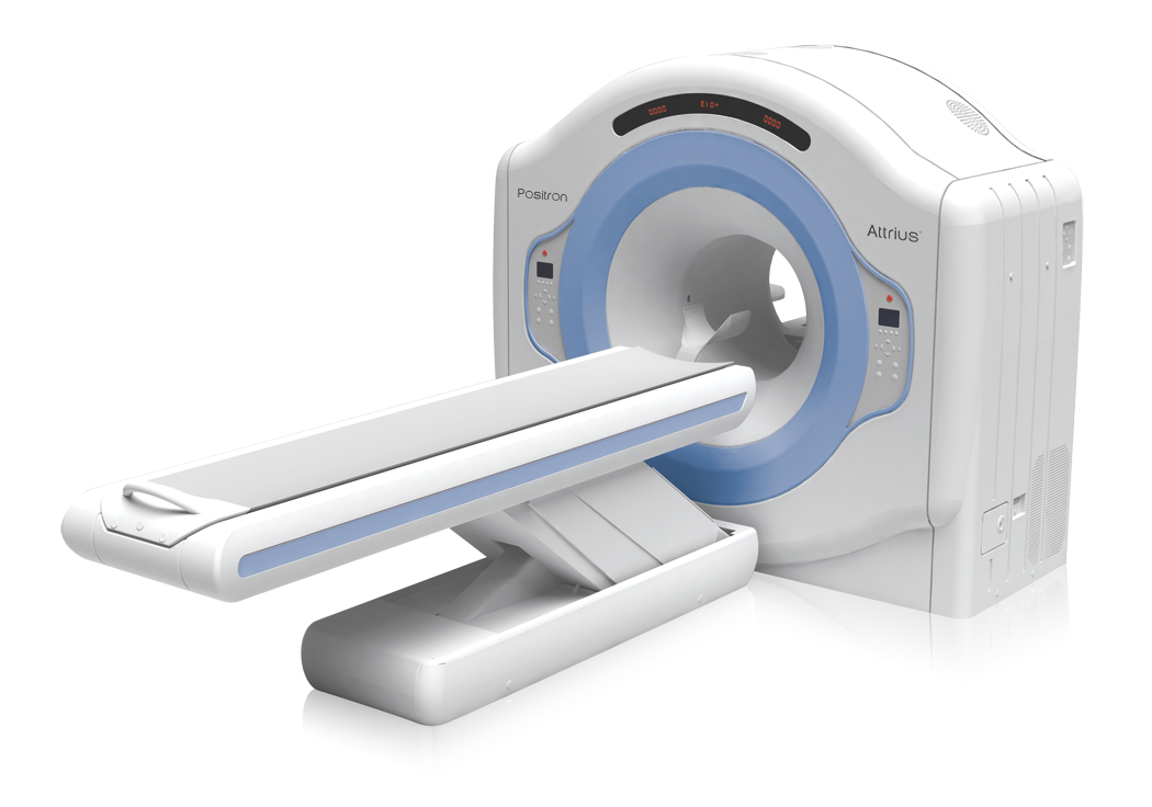
A PET-CT scan is an advanced imaging test that combines Positron Emission Tomography (PET) and Computed Tomography (CT) to provide detailed information about the structure and function of organs and tissues in the body.
A PET-CT scan is a diagnostic imaging procedure that combines two technologies—PET and CT—into a single scan. The CT scan provides clear images of the body’s internal structures, while the PET scan shows metabolic and functional activity of cells.
During the scan, a small amount of radioactive tracer is injected into the body. Cancer cells and other abnormal tissues often absorb more of this tracer due to their higher metabolic activity, making them easier to detect.
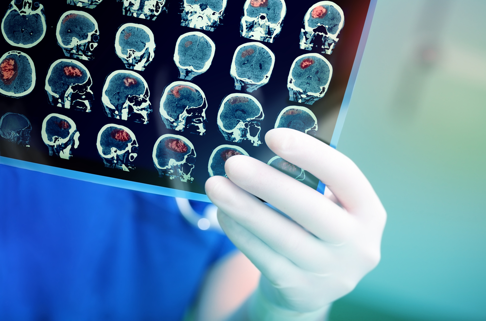
PET-CT scans are commonly used in oncology to detect cancer, determine its stage, assess response to treatment, and monitor for recurrence. They are also useful in evaluating certain neurological and cardiac conditions.
The procedure is non-invasive, safe, and usually completed within a few hours. Patients can typically return to normal activities shortly after the scan.
A PET-CT scan helps doctors accurately detect, stage, and monitor diseases by combining functional and structural imaging in a single examination.
PET-CT scans help in evaluating non-cancerous conditions such as infections, inflammatory diseases, and certain neurological or cardiac disorders by identifying abnormal metabolic activity in tissues.
In cancer care, PET-CT plays a crucial role in detecting cancer, determining its stage, evaluating treatment response, and identifying recurrence by highlighting areas of increased metabolic activity.


Preparing for a PET-CT Scan
- Fasting: Patients are usually advised not to eat for several hours before the scan.
- Medical review: The care team reviews medical history, medications, and prior reports.
- Tracer injection: A small amount of radioactive tracer is injected to highlight active tissues.
During the Scan
- The patient lies comfortably on the scanning table.
- The PET scan records metabolic activity, while the CT scan captures detailed images.
- The scan usually takes 20–30 minutes and is completely painless.
After the Scan
- Patients can resume normal activities immediately.
- Drinking fluids helps flush the tracer from the body.
- Scan results are reviewed by specialists and shared with the treating doctor.
PET-CT scanning is a non-invasive imaging technique that combines functional and anatomical information to support accurate diagnosis and treatment planning.
-
- Helps detect cancer at an early stage
- Provides detailed information on tumour activity and spread
- Assists in accurate cancer staging and restaging
- Supports treatment planning and response evaluation
- Non-invasive and painless imaging procedure
- Single scan provides both metabolic and anatomical data
-
- Low exposure to radiation
- Rare allergic reaction to tracer material
- Mild discomfort at injection site
- Not recommended during pregnancy unless necessary

• Combined functional and anatomical imaging: PET-CT combines metabolic information from PET with detailed structural imaging from CT, helping doctors understand both tumour activity and exact location.
• Early detection support: PET-CT helps identify cancer at an early stage by detecting abnormal metabolic activity before visible structural changes appear.
• Accurate staging and restaging: The scan assists in determining the extent of cancer spread and evaluating disease progression or recurrence.
• Treatment planning and monitoring: PET-CT is useful in planning radiation or chemotherapy and assessing how well a treatment is working.
• Non-invasive procedure: The scan is painless and does not require surgery, allowing patients to resume normal activities shortly after completion.